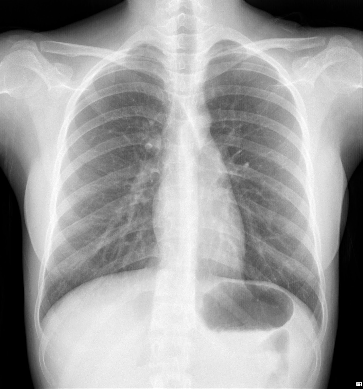
Confirm correct placement and position of the endotracheal tube, tracheostomy tube, chest tubes, central venous catheters, nasogastric feeding tube, pacemaker wires, intraortic balloon pump, Swan-Ganz catheters, and automatic implantable cardioverter defibrillator.Detect known or suspected pulmonary, cardiovascular, and skeletal disorders.Assist in the diagnosis of diaphragmatic hernia, lung tumors, and metastasis.Here are some of the reasons why a Chest x-ray is performed: Nursing Responsibilities for Chest X-ray.This diagnostic and laboratory procedure study guide can help nurses understand their tasks and responsibilities during a chest x-ray. Providing a calm and relaxed environment for the patient is indeed vital. In addition, producing a good quality image relies on the ability of the patient to cooperate, such as holding breath for a while. Nurses are responsible for ensuring the patient’s comfort while at the x-ray room since some may experience pain from injury or symptoms from a disease condition, as well as the apprehension about what the result may show. In the onset of the disease process of asthma, tuberculosis, and chronic obstructive pulmonary disease, chest x-ray results may not correlate with the patient’s clinical status and may even be normal. Rib detail images may be taken to delineate bone pathology, helpful when chest radiographs illustrate metastatic lesions or fractures. Expiration images may be needed to identify a pneumothorax or locate foreign materials.
#NORMAL CHEST XRAY FULL#
For critically ill patients who cannot leave the nursing unit, a portable x-ray machine is performed at the bedside using anteroposterior (AP) projections with an addition of a lateral decubitus view if a free flow fluid or air is suspected.Ĭhest images should be examined in full inspiration and erect if feasible to reduce cardiac magnification and demonstrate fluid levels. Other projections such as lateral decubitus, lordotic views, or oblique views can also be requested. A basic chest x-ray includes a posteroanterior (PA) view, in which x-rays pass from the back to the front of the body and a left lateral view. Air spaces normally seen in the lungs appear dark on the chest films. A chest x-ray is a painless, non-invasive test that uses electromagnetic waves to produce visual images of the heart, lungs, bones, and blood vessels of the chest. So a normal chest x ray, unfortunately, does not exclude many problems of the chest.Chest X-ray (Chest radiography, CXR) is one of the most frequently performed radiological examinations. This test is much better at detecting many problems like infections, cancer, clots, fluid and so on. This may include a Cat scan of the chest. I would discuss your symptoms with your provider and see what other testing is appropriate. I think that if you have symptoms referable to your chest, like a chronic cough or chest pain, and your chest x ray is normal, I would not necessarily assume all is truly normal. It is important that the radiologist reading your x ray has all your history and symptoms to get the most accurate diagnosis. If you do not bring in your old x rays, an abnormality may be not be seen or misdiagnosed as something concerning. Being a large patient may reduce the radiologist’s ability to see an abnormality. Sometimes the technique may not be the best. Sometimes it is hard to see and the radiologist may not report it. The reason for this is because the abnormality may be small or hiding behind another structure like the heart. There are also life threatening conditions that a chest x ray has no ability to identify like clots to the lungs or dissection of the aorta. Fluid around the heart and infections of the lungs be hidden. I have seen aneurysms of the aorta not show up.


In fact I have seen cases of lung masses and lung disease not clearly appear. In my experience a chest x ray does not show all problems but is a good start.


 0 kommentar(er)
